7 Free Neuroanatomy Midjourney AI images
Welcome to our Neuroanatomy image collection, featuring 7 free AI-generated images. This collection includes a diverse range of high-quality images such as stock photos, 3D objects, vectors, and detailed illustrations. All images are available for high-resolution download, and with our 'open in editor' feature, users can modify the prompts on the image detail page to generate personalized images based on their unique needs.
Related Tags
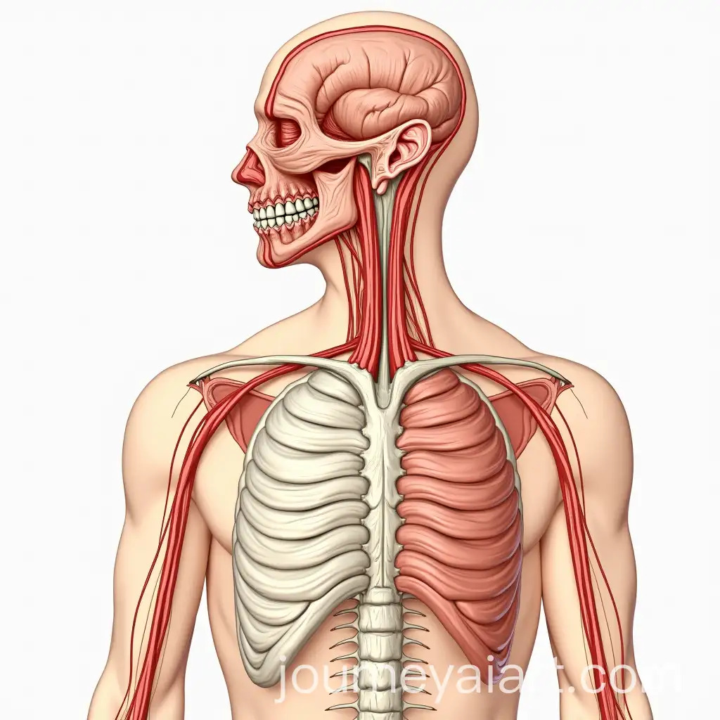
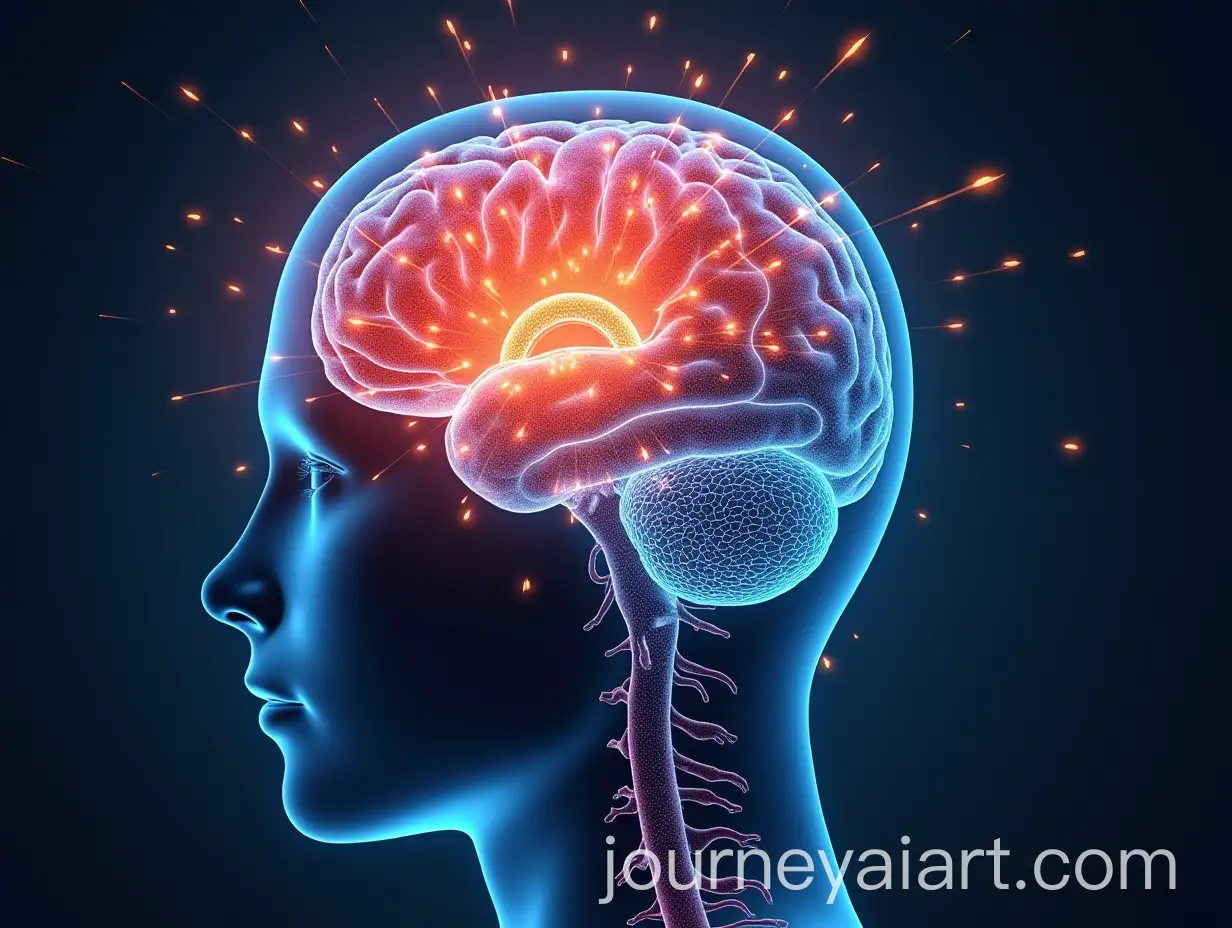
Neuroanatomy is the study of the structure and organization of the nervous system, with a focus on the brain and spinal cord. It is a crucial area within neuroscience that helps researchers and medical professionals understand how different parts of the brain and nervous system connect and interact. AI-generated images in this category can depict intricate neural networks, brain sections, and other detailed representations, making them valuable for educational, scientific, and medical uses.
Understanding Neuroanatomy: A Key Area of Brain Science
Images related to Neuroanatomy are widely used in educational resources, healthcare, and research. Medical students and professionals often rely on visual aids such as detailed diagrams of the brain, neural pathways, and synaptic connections to learn or explain complex concepts. AI-generated images provide precise, customizable visuals that can enhance teaching materials, presentations, and patient communication in fields like neurology, neurosurgery, and psychology.
Applications of Neuroanatomy Images in Education and Healthcare
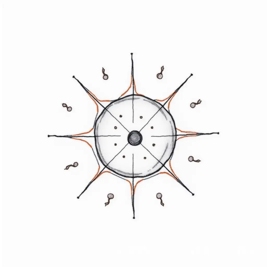
Creating AI-generated Neuroanatomy images is easy with the right prompts. Start by describing the specific brain region or nervous system component you need, like 'cross-sectional image of the cerebral cortex' or 'neural pathways in the spinal cord.' Once generated, users can refine details, such as the addition of labels or annotations, to ensure the image meets their requirements. The 'open in editor' feature allows further customization, so anyone can craft professional-quality neuroanatomical visuals with minimal effort.
Creating Neuroanatomy Visuals: A Step-by-Step Guide for AI Enthusiasts
The integration of AI in neuroanatomical studies is accelerating advancements in how we understand the brain. AI-generated images allow for faster, more precise visualizations, which aid in research, diagnostics, and education. In the future, AI may be used to model neural networks in real-time or generate personalized visuals for patient care, revolutionizing the fields of neuroscience and neurology. As AI continues to evolve, its applications in neuroanatomy are expected to grow exponentially.
Future Trends: How AI is Shaping the Study of Neuroanatomy